Museums theme – making Split + Splice: Fragments from the Age of Biomedicine
Article DOI: https://dx.doi.org/10.15180/170813
Abstract
The wide-ranging personal experiences of medicine that people bring with them on their visits to medical museums arguably give medicine an advantage over the histories and material culture of other technologies and sciences. No other science has ‘content’ that is held quite so deeply inside the visitor’s conscious substance and psyche. But if mis-handled through sensationalism, this can easily backfire, catapulting the visitor into a state of fear and abjection. This article outlines a sustained and award-winning temporary exhibition project that aimed to engage visitors by locating them in the engine room of biomedicine rather than the operating theatre. Four post-doctoral historians of contemporary medicine and a creative director worked together with exhibition designers in a research context to show how epistemology can be dovetailed with aesthetics to bring theory and practice together for a wider public at Medical Museion, Copenhagen.
Keywords
Biotechnology, Dibner Prize, exhibition design, exhibitions, Foucault, Interdisciplinarity, medical humanities, Museum Visitors, Personhood, Research-led Museum Practice
‘Technologies of the self’ at the Medical Museion, Copenhagen
https://dx.doi.org/10.15180/170813/002Split + Splice: Fragments from the Age of Biomedicine took place at the University of Copenhagen’s Medical Museion from June 2009 to April 2010. It was created over a three-year period by a dedicated, interdisciplinary and international team of curators, designers, and museum staff, supported by a range of professional colleagues from the academy, biomedicine, fine art and more.[1]
In this short overview of considerations concerning the development of the exhibition and the impact that it had on visitors, I outline, as the exhibition’s Creative Director, some of the significant intellectual and practical aspects of the exhibition’s innovations. Split + Splice took place in the overlapping contexts of twentieth and twenty-first century medical humanities research, and the experimentation that is possible in university museum environments. Having set the scene, I will then look at the relationship between theories of personhood and flesh-and-blood museum visitors. Theatrical strategies of distanciation will be shown to be of considerable assistance in accommodating the visitor in medical museums, and we will see how down-to-earth Foucaultian concepts of power can be when they are applied both literally as well as figuratively to displays of medical technologies. In the second part of this paper, I go on to recount the ways in which the exhibition team productively meshed methodologies and skills through brainstorming and iteration, and how we moved beyond narrative as an organising principle for exhibition experiences.
Increasingly over the past decade, rapprochements between higher education establishments, research centres, and heritage and collecting organisations have highlighted what is shared by all of them as ‘knowledge institutions’. This is leading to deeper engagements with material culture on the part of academics, more intellectually robust and exploratory exhibitions, and the emergence of new methodologies in both the humanities and in museum practice. Director Professor Thomas Söderqvist of the Medical Museion worked extensively with staff and colleagues to create a research-based institution which experiments and innovates:
By focusing on integration between research, collecting, education and dissemination beyond the museum world, the Medicinsk Museion will go beyond the traditional division between universities as pure research-and-teaching units, and museums which are primarily collection and dissemination institutions.[2]
Split + Splice was the first major such project at Museion. Its main funder was the Novo Nordisk Foundation, through the overarching research award Danish Biomedicine: 1955–2005: Integrating Medical Museology and the Historiography of Contemporary Biomedicine, for which Professor Söderqvist was the Principal Investigator.[3] The exhibition was the flagship of a sheaf of impressive ‘outputs’ associated with this pioneering research project.
The exhibition itself was a multi-media yet low-tech project, which brought together a wide variety of materials, content, ideas, instruments, collections and disciplines in entirely new ways. It was not just about the science of biomedicine: it was about the cultures of biomedicine and the kinds of societies that its practices produce. It was about the inter-relations between biomedicine and the complexities of twenty-first-century living, addressing the long history of modernity that is present in biomedical technologies. Each of the concept-based displays meshing both historical and contemporary objects grew directly out of extensive curatorial field research into the history of recent biomedicine coupled with epistemological inquiry into both effects and affects of biomedical practice. With Split + Splice, we took the visitor beyond the ‘user end’ of biomedicine and into its engine room, bringing some of the big ‘invisibles’ of biomedical practice to a larger public in innovative ways (Fleming, 2007). Questions of samples and storage, data generation and management, the integration of analytical instruments with research and clinical bureaucracies, and the legal frameworks of biomedicine were all on our agenda.[4] These are critically important practices and yet they are also very complex to materialise and to visualise. They are not at all ‘visceral’ – they are unlike anything a visitor might have come to expect from a medical museum. And yet this exhibition experiment elicited deep reactions from its audience by engaging them in a conceptual inquiry about biomedicine today, taking people and biomedical technologies out of the hospital and bringing them together again entirely reconfigured inside an arena of thought.
In the realm of museum display, the history of medicine arguably has an advantage over the histories of other sciences. This ‘advantage’ is also among the most difficult to deploy, the most volatile, and the most easily misused by exhibition makers. It is the advantage offered by the wide-ranging personal experiences of medicine that visitors bring to the museum, and the myriad ways in which those visitors’ identities are a priori entwined and fused with their experience of their bodies and their bodies’ health. No other science has content that is also held deep inside each and every museum visitor’s conscious substance and psyche.
Like a built-in counter-weight, this ‘advantage’ of the subject matter of medical history exhibitions ties the visitors’ fascination with the exhibition to their sense of self. It is a tethering that, if mis-handled, can just as easily backfire – catapulting the visitor into a state of fear and abjection, and casting the exhibition (and its museum) into a no-go zone. Sensationalist misuse of instruments such as surgical saws, needles, laparoscopes, and specula – all of which enter the body – or of full-body MRI scanners, early X-Ray rooms, and covered stretchers – all of which enclose the body – are among the prime examples of medical exhibition shock tactics that can badly rebound.
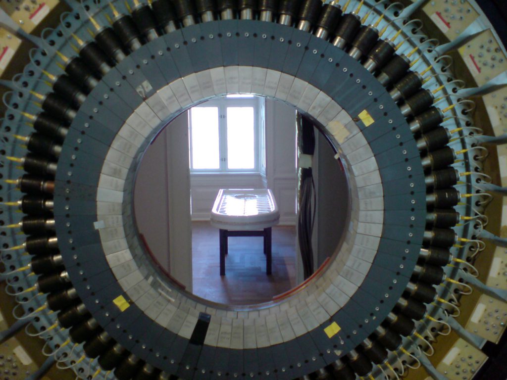
One way to avoid this rebound is to look at the very relationship between medical subject matter and personal visitor experience as part of the exhibition itself; a philosophy lesson from the inside out, with help from the Brechtian distanciation effect (Pramaggiore and Wallis, 2005).[5] This is what we set out to achieve with Split + Splice. As historians and thinking museologists, many of the tools we have with which to attempt to effect this were first given to us by Foucault. Conversely it is not surprising that this great historian of modernity would first have looked to histories of medicine, psychiatry, the body and sexuality: this nexus contains so very much of all that it is important to reflect upon. And it is central to understanding not just biomedicine but also how people engage with cultural activities such as museum exhibitions.
In an anthology of essays such as this, wishing to investigate and problematise the oft-simplified relationship between history of technology and history of science, we can have helpful recourse to one of the many amazing tools Foucault wrought for us. His theory of ‘technologies of the self’ was generated from within the particular frame of his research that could be described as history of medicine writ large. Technologies of the self engage variously with the other three technologies that Foucault also identified as part of four interlocking systems: technologies of production; technologies of sign systems; and technologies of power. Technologies of the self ‘permit individuals to effect by their own means or with the help of others a certain number of operations on their own bodies and souls, thoughts, conduct, and way of being, so as to transform themselves in order to attain a certain state of happiness, purity, wisdom, perfection, or immortality’ (Foucault, 1988). But this of course never happens in isolation. When these personal acts of self-realisation come in contact with what Foucault called technologies of power – which compel people to internalise attitudes and beliefs that are alien to them via subtle coercion and social pressures – those technologies of the self that originate with the intent to improve one’s lot can become enmeshed in other acts and actions that can actually be compromising for the realisation of self. Yet these compromising ‘governmental’ acts that issue from the internalisation of beliefs alien to the self can at times present themselves as the only way to realise a self, often presenting themselves so invisibly as to appear even to be the best way to realise a self (Foucault, 1988).
With biomedicine, many scientific and social changes and exchanges converge in the human body itself, and are therefore difficult for individuals to observe with any distance or objectivity. But what if we were to examine, in an exhibition, this ‘contact’ between the patient and the biomedical apparatus in all its complexity? Contact that is conceptual as much as physical; contact that takes place sometimes even at a distance, unwittingly, and at times without informed choice or consent. If we were to create an exhibition to unpick this diffuse and decentralised contact, bringing together people and instruments into an arena of thought, we would have before us the panoply of technologies identified by Foucault:
technologies of production – instruments of analysis and computation;
technologies of power – regimes of medical authority and knowledge;
technologies of sign systems – medical vocabularies and indexical relations;
technologies of the self – in which the individual attempts to care for the self, in part by engaging with all these other technologies.
This complex of technologies is, in essence, the scenario which the core exhibition team for Split + Splice attempted to both deconstruct and render visible – not for the viewer, but around, through and with her – addressing her as a thinking equal, not as a patient-in-waiting. Though we did not formulate the exhibition directly as a Foucauldian project, this description of our project is accurate, and speaks to the permeation of Foucault’s thinking through much history, historiography and philosophy of science, as well as in radical museology and critical representational practices. Constructing an exhibition that deconstructed this patient/biomedical-apparatus ‘contact’ zone functioned to introduce an empowering distanciation at the core of the exhibition; it thus enabled us to deploy the powerful draw of medical subject matter in an exhibition without wilfully abjecting the visitor.
Proposing implicitly that the visitor is among other things a delicate instrument and a deployer of ‘technologies of the self’ put the visitor at the heart of the exhibition whilst concurrently questioning the very meaning of the term ‘technology’ in a biotechnological age. The exhibition both acknowledged and highlighted visitor subjectivity by placing the visitor ‘inside’ an arrayed mechanics of biomedical practices, and yet resolutely positioned them outside of biomedical treatment.
We wanted to address the social, political and cultural enormities of contemporary biomedicine without losing sight of the historical. As historians and epistemologists, we did not want to teach people the science, but rather to teach people how to think about science; to think about how bodies are contingent, flexible, fluid, resilient; how materials, tools and instruments have a history; how conditions for the production of medical knowledge change over time.
This last is of most interest in the context of this anthology: engaging with an expanded field of conceptual and theoretical ‘technologies’ as well as physical technologies enabled us to transcend the reductive, causal, blandly narrative threads often constructed in museums between science as a practice and the material culture of technology.
Of course, this is more easily said than done. It was important to us to get beyond ‘stories’ altogether, avoiding the built-in tendency of narrative to require both a neat ending and a display-case-bound object-anchor as illustrative evidence with which to tautologically legitimate itself. We then had to find ways to distinguish between different categories of object and of concept without inadvertently hierarchizing them. We set ourselves a tall order.
In what follows, I will outline some of the process which produced this ground-breaking result: an experimental exhibition which won the 2010 Dibner Award for Excellence in Museum Exhibits of the Society for the History of Technology. I will show how our conceptual approach led us to define unusual criteria for exhibition-making. Along the way, I will identify some of our creative techniques, and address some of the object choices we made.
Caveat lector: this short account can be neither comprehensive nor strictly developmentally chronological (Fleming, 2010; Fleming, 2013).[6] The process of creating this exhibition involved the pooling of knowledge and the transference of apparently disparate skills across a small, highly trained group; skills some of which in themselves had already taken decades to acquire.
This ‘skills exchange’ involved history of medicine and epistemology of science practice, history of material culture, exhibition-making, collection management, design and more. In this crucible, we also forged new ways of working that will inform all our work into the future. The idea was to produce a double-edged tool which would help us create the exhibition, a tool in which epistemological and aesthetic enquiries would function in equal measure and in tandem: we wanted to deploy aesthetics as an analytical instrument as well as a communication tool, and we wanted to show that epistemological inquiry can guide what an exhibition ends up looking like. For example, an epistemological enquiry about standardisation in laboratory practice can uncover some of the reasons for the ubiquity of the appearance of plastics in recent biotechnology. In a complementary fashion, paying critical aesthetic attention to these plastics can contribute to the granularity of that epistemological research: colourways in the plastics relate to workflows in the lab; transparent high optical quality plastics are created for spectroscopic scanning with new analytical instruments. Because aesthetic judgements and choices are as significant in the production of objects and meaning as they are in their reception, aesthetics can be employed analytically. Further, in creating an exhibition, one uses aesthetic judgement in designing the display itself in order to bring to light the deeper-level functions, clues and meanings of the very objects one is displaying.
A series of team workshops or ‘brainstorms’ between myself as Visiting Associate Professor and the four post-doctoral researchers who were to co-curate the exhibition with me were held early on in order to identify the overarching issues and themes shared across their widely diverging fields of research.[7] None of them had ever created an exhibition before: as Creative Director, I gave a number of crash-course lectures in museum practice and exhibition-making which were essential to the production of the exhibition – such as Exhibition Making Process, Project Management for Exhibitions, Deriving Workplans from Concepts, Writing for Exhibitions, Artists Working in Science Museums, Working with a Designer, and so on. Further, in a two-day design workshop with UK-based exhibition designer Calum Storrie, we explored the spatialisation of thought, and ways in which the galleries of the Museion could be used conceptually rather than consecutively – working against the temptation to express concepts and ideas through storytelling.
By mid-2008 we had created a framework (including an Exhibition Brief, a list of agreed Limit Parameters or creative constraints, and other process-oriented guidelines) with which to begin the conceptual materialisation of the exhibition itself, and to choose exhibition objects from both Museion’s collections and further afield.
Our Exhibition Brief stated concisely that: ‘The core aim of the exhibition is to facilitate visitors’ informed reflections upon the ways in which recent biomedicine challenges significant cultural categories including the body and identity, therefore influencing our very understanding of ourselves as human beings, our sense of “personhood”. The exhibition will explore how both identities and bodies – two sides of the same “personhood” coin – are produced, constituted, re-engineered, collected, circulated, fragmented, hybridized, mediated, suspended, digitized, controlled and categorized by current biomedical practices.’[8]
The parameters and creative constraints we gave ourselves within which to work were refined over the conceptualisation period, and yet remained very close to some of those we first set out with, such as:
Every object and image must relate to more than one issue, so there will be no one-to-one equivalences between idea and image: everything needs to do the work of problematising several issues and practices at once. Everything we do must help to destabilise the truth-value of representation, particularly forms of realism, so that people can by extension have a way to question both scientific objectivity and transparency as a metaphor for intellectual and social clarity. [We will] introduce both historical and representational critique and reflexivity into everything we do.[9]
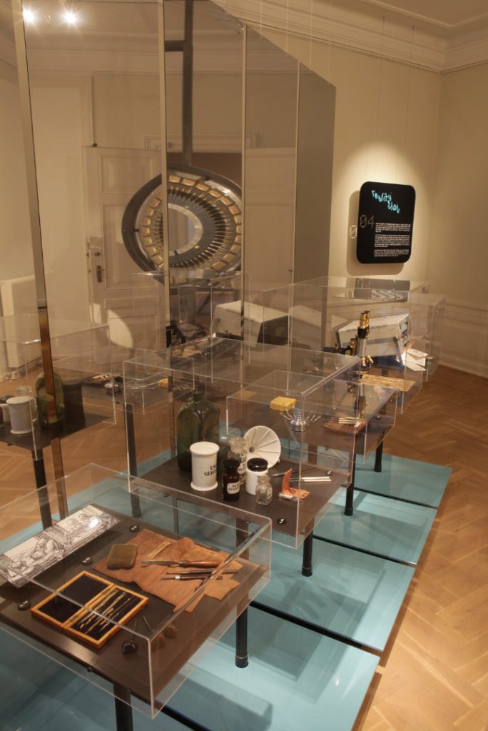
One example of the meshing of representational and historical reflexivity and critique in Split + Splice can be seen in Section 4 of the exhibition, entitled Reality Show. Instruments and accoutrements from five different historically-bound techniques for representing the human foetus were explored: Drawing and Engraving; Preservation of Specimens; Wax Models; Histology; and Ultrasound. These five cases were set in front of a two-way mirror which visitors could step behind. On the other side, they found in isolation the realistic images which these complex techniques were capable of producing – a technical complexity often subsumed in realism, a genre which borrows the appearance of life, creating images easily press-ganged into serving as truth claims.
It is worth noting that with the Museion Vision for an integrated collection and research institution, Director Söderqvist had charged the post-doctoral researchers with collecting the material culture of contemporary biomedicine as part of their field research process. These new collections were a starting point for the development of the exhibition – both the objects and the problems they presented. The huge ‘invisibles’ of contemporary biomedicine and its complex practices would be difficult to represent with the mainly standardised disposable plastic materials of contemporary biomedicine which had rightly been amassed by the post-docs. Many of these objects were both anodyne and existentially complex: how to make the latter shine through the former? Because material gathered for collection purposes is very different from material needed for an exhibition – which requires contextual, correlative and other objects to create counterpoint in order to inspire both understanding and excitement.
From our Exhibition Brief again:
The exhibition medium is an environment: this environment needs to embody the contexts of contemporary biomedicine, not just ‘explain’ these contexts didactically. Visitors may encounter refrigeration units and biosecure areas, […] incubation units for animals and unusual electronic and mechanical instruments.
- the audience’s own bodies will be used as ‘instruments’ by which those same individuals can ‘measure’ realities of contemporary biomedicine phenomenologically;
- the in-house collections at Medical Museion will be used in analogical, material and metaphorical ways as well as historical ways;
- the exhibition will use medical instruments (research and clinical) in non-medical ways that open them to critique and playful exploration by the visitors;
- simple mechanical instruments from medicine and everyday life which are well understood will be used as a springboard to explore electronic and digital aspects of biomedicine: a spinning salad dryer can help in understanding a centrifuge.[10]
With these ideas among others to the forefront, we began to look closely at the Museion collection, the materials that the post-docs had collected, and other objects – including some that were very large indeed – which were further afield and embedded in life sciences research labs, hospitals, and clinical environments. With the extended community – and inestimable help – of the University of Copenhagen’s Faculty of Health and the contacts the post-docs had made during their field research, we were also able to call on a much wider base for exhibition objects. We moved from ideas to objects and back again quickly and often, during a process of rolling brainstorms which often involved the objects themselves.
In all, and including the design phase, there were some 45 brainstorms, workshops and other concept meetings and seminars which formed the developmental arc of the exhibition. It was during these sessions that we were able to dovetail aesthetics and epistemologies of biomedicine via object and design choices. The structure of the brainstorms themselves was fairly standard: identification of issues to be explored, preparation and pre-circulation of a framework for the discussion, an exploratory and open meeting of finite length which is whiteboarded by the discussion leader, post-meeting analysis and capture of the outcomes which is circulated, and finally the incorporation of those outcomes as part of the givens for future brainstorms. For example, having held several brainstorms about medical visualisation and about data management and modelling in medicine, we then held a brainstorm about overlaps and connections between these two intertwined fields.
A key point of the success of these discussions was that we were in fact not only interested in each others’ work, and very different from each other in terms of our knowledge bases and fields of practice, but we were also dependent upon each other for pieces of the puzzle. Without taking on trust each others’ work and opinions, we would not have been able to move forward to create an exhibition. So the oft-heard brainstorm technique rule of suspending judgement also meant, in this case, maintaining faith in each other’s judgement. In spite of the scheduling pressures we experienced, the time we spent on conceptual work towards the exhibition was not only essential, but an exciting arena of sharing knowledge and finding sound conceptual links between fields of study, representational and aesthetic structures, epistemological analyses, collection objects, urban and exhibition spaces, and visitor experience from a phenomenological point of view. We had to move quickly, be concise and rigorous, and remain open – and as relaxed as possible. This last was of course the most difficult.
To develop skills of ‘thinking through objects’ in the historians of the group, we held a number of seminars to examine in depth the materiality of the Museion collections, some with the precious contribution of Collection Manager and Conservation Director Ion Meyer. Other such seminars were those in which collection instruments were used to ‘play with’ – both as interactives and as metaphors. Jan-Eric Olsén, whose work focuses on medical visualisation, gave an instrument-rich seminar tracing the continued presence of the precepts of Renaissance optics inside the high-tech imaging of contemporary biomedicine.
As a co-relative to the history of optics in medicine, we turned towards biomedicine’s observation techniques and technologies, examining the centrality to both museums and biomedical practice of the importance of display; a key crossover that we had identified as both epistemologically significant and highly useful to us in an exhibition context.[11] The fine line between attentive care and medical surveillance was explored in relation to the formation of the body politic through, among others, public health policy and data collection: once again there were synergies with museum practice – collections held in trust for the nation and surveillance cameras as a form of ‘security’.
A number of research outings were also significant. We went to the Copenhagen City Museum with a view to critiquing object choices, exhibition design, display texts and more. We were particularly impressed by the exhibition Storbystrømme, concerning underground waterways and electricity cabling, which was designed by Mikael Thorsted. This honed the capacity of the historians to articulate what works in museum displays and why. Susanne Bauer followed up a contact at the Danish Data Historical Association which led to an Aladdin’s Cave of computing history: the Association became a major lender to the Data Avalanche section of the exhibition. Søren Bak-Jensen crucially arranged for us to visit the Cold Rooms and refrigeration team of the University’s Panum Institute, as well as to take a behind-the-scenes tour of the High Court building immediately opposite the Medical Museion. This very, very large ‘object’ – visible from a number of the galleries of the Museion in which Split + Splice was to take place – is also the site of the arbitration of legislation in relation to biomedicine. This took to a new level an awareness of the position – both literally and figuratively – of the Museion building, which had been the College of Physicians and Surgeons (founded 1792). Architecture in the dense power-centre of the city is an intersecting set of signs – each ‘technology’ visible as an edifice to the other through overlooking windows.
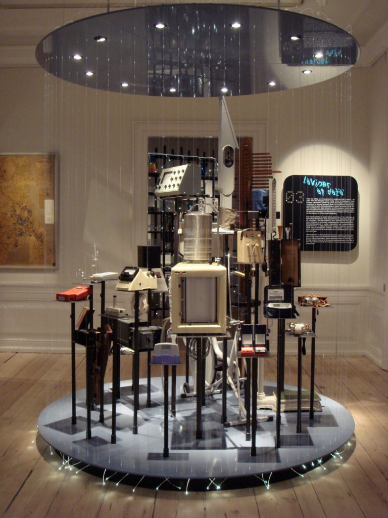
Indeed, we even workshopped subjects which we did not foreground in the exhibition. One workshop in particular, entitled ‘Hope and Health: The User End, Biomedical Benefits’ was particularly helpful in that the discussions made clear the fact that this kind of ‘patient-focused’, emotionally charged content would be exactly the sort of material that would appear to meet visitor expectation of our exhibition, whilst in actual fact eliciting visitor anxiety and inexorably drawing the project towards presenting a view of biomedicine reduced to a narrow interpretation of progressivist ‘public service’. Which is not to say that biomedicine does not produce huge advances in human health, only that this was not the subject of the exhibition we were making.
The Museion collection itself, which we wanted to engage as a set of historical anchors for the exhibition, is as impressive and as frustrating as any university museum collection can be. Universities the world over have an urgent responsibility to release the huge interdisciplinary research potential locked up in their museums, and which is currently often caught between underfunding of collection management and overuse by the untrained.[12] In spite of the highly skilled and dedicated Museion staff, working far beyond the call of duty, the exhibition team had difficulties in locating materials we were interested in, both through the range of different catalogues and in visits to store-rooms scattered about the University of Copenhagen.
But serendipity can be an important research tool – even in the best-endowed of museums. In certain cases, unique elements of the Museion’s exciting collections were a focus for particular brainstorms: an important donation from the DAKO company (a diagnostics manufacturer of antibodies, reagents and instruments) includes the original materials used by founder Dr Niels Harboe when he was the first Director of the Protein Laboratory of the University of Copenhagen in the 1970s. Among these objects are the simple tools used by Harboe to produce antibodies in rabbit blood for harvesting and use as a diagnostic tool in human illnesses. With the exception of the DIY electrophoresis machine, all the instruments could have been found in the visitor’s personal home: sieves, vodka bottles, bottle capper, funnels, and more. In the exhibition, they provide a familiar cultural link to biomedical practice, and a bridge for the understanding of a wider public – a public we hoped to see coalesce before us through self-selection.[13] Extending this bridge, we displayed two live rabbits alongside this material: these were not lab animals, but pets. The intent was to bring human-animal relations into a kitchen-like context that invited visitors to relate the experience of pet-owning with issues of animals in biomedicine: distanciation can paradoxically bring one closer to the philosophical issues. Resolving this section of the exhibition involved some of the most complex of our discussions.
And what of illuminating the epistemological and existential importance of the more mundane accoutrements of biomedicine? A particularly significant object which helped us begin to resolve this problematic had been collected by Sniff Andersen Nexø, whose research has focused on fertility and its technologies, foetuses, abortion and the social structures that create these practices and subsequently are created by them. An entirely non-descript plastic receptacle, only some ten centimetres tall, is a container for tissue which is the disposable part of a clinical abortion apparatus.[14] This container is a mobile component of a biomedical technological device, and it partakes of the degree of standardisation and industrialisation of medicine which is a hallmark of biomedicine today. This container in turn is also harnessed to a ‘technology of power’ – of legislation, for example, in relation to the European Union Tissue and Cells Directive. Concurrently, it is enmeshed in a contested field of social interactions which include notions of personhood.
It became clear in discussion that the container itself in some social contexts (such as debates concerning the dates at which life is believed to begin after the moment of conception), is made to perform the inadvertent function of ascribing personhood to its contents, independently of the medical and legislative structures in which its use is set. Thus, this simple plastic object is actually incredibly volatile socially and has significant roles which it plays. In the highly contested arena of abortion, for some it ‘contains’ human remains, for others waste tissue. The very contour of the container itself seems to create the simulacra of personhood onto which is subsequently grafted a myriad of social, ethical and political projections.
From the brainstorm discussion about this object came the realisation that all biomedical ‘containment’ and containers are a mechanism of the type of instantiation of personhood that we wished to address. This simple link became one of the core organising principles and leitmotifs of the exhibition through which we were able to connect directly to the visitor; the visitor for whom personal integrity might well be experienced – for better or worse, rightly or wrongly – as residing in the fragile impermeability of his or her liminal bodily contour.
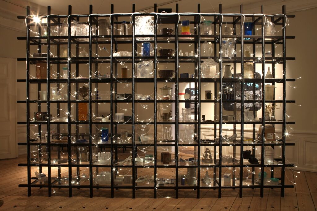
We began to look more closely at other biomedical containers from this point of view, to see how they might, within their delimited and defined ‘spaces’, similarly configure the ambiguities of various categories such as ‘life’, ‘biohazard’, ‘genetically modified matter’ and so on. Going from older and bigger hospital ceramics to newer and smaller containers for biomedical sampling of increasingly minute fragments of biological matter could also serve to trace the historical origins of these contemporary practices in earlier, larger-scale anatomical dissection. Containers also imply flow – flows of liquids from one container to another, flows of data from analysis of contained biomaterials, biomedical workflows, cashflows of biotechnology corporations.
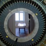
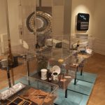
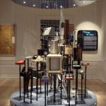
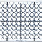
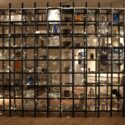
Because there has always been ‘flow’ in medicine, and containers to contain it. Museion’s collection is rich in the stuff of medical history from Enlightenment on, and we chose a wide range of containers of varying materials, histories and use – for drugs, samples, serums, data and more. We used this material both historically accurately and metaphorically as an aid to highlighting the less visible, more conceptual ‘flows’ of contemporary biomedical processes, and we displayed it in a structure inspired by another apparently mundane object: the ubiquitous 96-well plastic microplate.
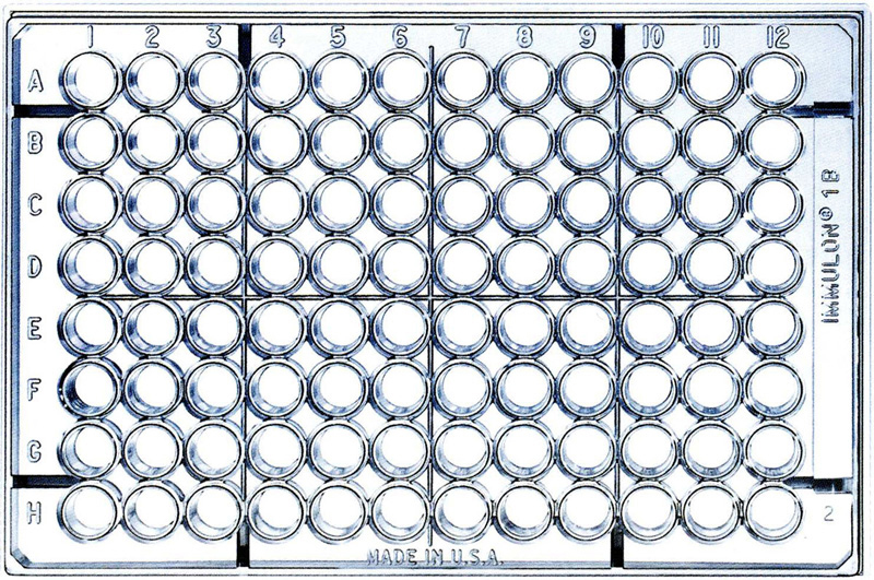
This plastic container, each well of which functions like a tiny test tube, has an alphanumeric grid for sample tracking, high optical quality plastic for throughput scanning, is stackable and storable hot or cold, radiation sterilisable, amenable to manual or machine manipulation and filling, can be heat sealed with foil or film, and is a central part of all research fields in life science.[15] As Susanne Bauer pointed out, its grid mirrors that of the punch card, and it is designed specifically to enable the tracing of digital data generated from the analysis of the biomaterials contained within each of its identifiable wells. It is a ‘technology of production’ of the first order: an instrument of analysis and computation that produces particular biological ‘bodies’ alongside its data ‘corpus’.
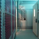
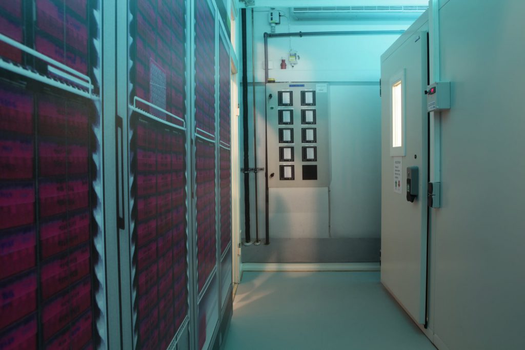
Following a set of brainstorm meetings about the use of temperature in biomedical practice, which ranged from discussions about the PCR machine through to IVF incubators and cryogenics in biobanking, we realised that a laboratory Cold Room was a ‘container’ large enough to encompass even our visitors, and to give them a sensory understanding of not only the conditions of biomedical practice, but also a visceral experience relating to biosample storage. The Cold Room which we installed as part of Split + Splice was not only an actual instrument but also a phenomenological experience. It became one of our major exhibition objects, and a complex project management segment of the exhibition creation. Without the ingenuity of our Curatorial Assistant Jonas Paludan, its inclusion would not have been possible, and a core through-thread would not have had its major anchor.
So we chose objects, yes; but also temperatures, sounds, textures, films, animals, cameras and more. Of course, object choices are also intimately related to design problems, and we dovetailed our design process with our object selections – sometimes out of necessity rather than choice – and were very lucky to have been working with exhibition designers Mikael Thorsted and Lars Møller Nielsen of Studio 8. The brainstorms and workshops which we held with our designers were no less intensive than those we had held as a core team. Fortunately they were interested in being so closely involved in the devising process of the exhibition, and the result showed their commitment to the project. Once again, knowledge pooled was also skills exchanged, and the time taken for exploratory discussion was rewarded by a high degree of innovation.
Some of the issues we outlined to them in our Design Brief indicate the integrity of design with object choice and concept:
The biggest problem is how to create overall exhibition coherence without falsely totalising the information within it. Everything in the exhibition must work towards grounding the visitor’s body in their own senses, their phenomenological experience, and their subjectivity so we can get straight inside them – YET everything in the exhibition must work towards showing them how much their bodies are fragmented and displaced and reconstituted by biomedicine. A whole body is required in order to sense (and thus cognitively recognise) the body-in-pieces. […] We want to use objects as if they were explanatory texts for each other.[16]
This relationship between fragment and whole – both in the display itself and in the visitor experience – was ultimately rendered in a careful balancing act of contextualisation that kept objects and labels apart in the exhibition, and therefore invited the visitor to observe relations between things. We were asking them to hypothesise themselves, to draw their own deductions from our constellations of biotechnological instruments, consumables, techniques, processes, products, affects and more. We did everything we could to foster self-reflexivity on the part of the visitor: awareness of self as a sensing instrument, as a body moving through space, as a body with integrity at a large scale, as a body with a unique identity.
Visitor flow, which had already been problematised in the workshops we had held early on with exhibition designer Calum Storrie, came into stark focus at this point. The grid of the gallery spaces and the sketch-up software used in the design process came out to meet the grid of the epidemiological computer punch card and of the plastic microwell. It became clear that there was a structural similarity to the ways in which the visitor would circulate in the exhibition and the ways in which biomaterials and data of all kinds circulate within biomedicine. This structural similarity of ‘flows’ became a foundational tenet through which we were able to display, alongside many objects, the very instant of engagement between a ‘technology of the self’ and a ‘technology of power’. At any given moment in the visitor’s flow through the show, there was a concurrent instance of flow within a biomedical process to be pondered.
In the strictly ‘hardware’ sense of the term, technologies are pivotal parts of processes – scientific and otherwise; technologies are both produced by processes and produce them. The reduction of technology to hardware which is in part unwittingly occasioned by museological focus on collecting objects rather than concepts has meant that instruments – biomedical and otherwise – are often the focal point of exhibitions which consequently explain the world in instrumentalist ways. When this is the only ‘story’ that is told, then only part of the story is being told. Split + Splice, and the way we constructed the exhibition, has shown that instruments can be employed in ways much more complex, metaphorical and allegorical; ways that point the visitor to the concepts and the processes that swirl around and through these technologies; ways that show the visitor the larger picture – of technologies of the self, of power, of signs, and of production; production that has produced the very ‘hardware’ in the display case before them. It is worth remembering that an exhibition, too, is a technology.
Tags
Footnotes
Back to text
Back to text
Back to text
Back to text
Back to text
Back to text
• ‘Biomedicine and Aesthetics in a Museum Context’, a three-day closed workshop bringing together over thirty biomedical practitioners, medical historians, philosophers of science, artists, exhibition designers, material culture experts, etc.
• ‘Biomedicine and Art’, a one-day public conference collaboration with the Royal Danish Art Academy showcasing exhibition makers, art historians, artists and biomedical practitioners who work with artists. Back to text
Back to text
Back to text
Back to text
Back to text
Back to text
Back to text
Back to text
Back to text
Back to text


